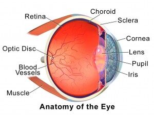
Retinal Anatomy
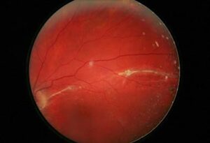
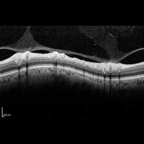
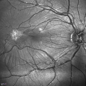
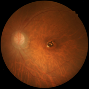
The retina is the light-sensitive layer of the back of the eye that makes focused light into an electrical signal that is carried to our brains through the optic nerve. Light travels through different parts of the eye to become focused light on the retina. The cornea, iris and lens are in the front of the eye and help to focus the light. The center part of the eye is made up of a clear gel called the vitreous (vi-tree-us). The vitreous is attached to the retina. Sometimes the gel forms clumps, called floaters, that cast shadows on the retina which are seen as dark specks in our vision. These changes in the vitreous often lead to the gel pulling away from the back of the eye. When this happens it is called a Posterior Vitreous Detachment (PVD). If the gel pulls too hard on the retina, you can notice flashing lights. Rarely, the vitreous can pull hard enough to create a small retina hole or tear. Fluid may pass through the retinal tear and lift the retina off the back of the eye and create a retinal detachment. This will often be noticed as a dark curtain in your peripheral vision. A retinal detachment can cause loss of vision if left untreated.


